What Does Skin Cancer Look Like on the Face?
Niche: Health & Medicine Content Type: Topic Explanation Why It Matters: Skin cancer is a global health issue (especially melanoma), and this query is critical for early diagnosis in any country.
How Skin Cancer Looks on the Face: A Complete Visual Guide
Skin cancer on the face can look like a harmless mole, a pimple that won't go away, or even a small scar. The face receives the most sun exposure over a lifetime, which is why up to 80% of all basal cell carcinomas occur here.
But there's no need to panic. Most spots on the skin are benign. This article aims to teach you how to distinguish warning signs from harmless changes.
The Bottom Line: What You Need to Know First
There are three main types of skin cancer, and each looks different.
Basal Cell Carcinoma (BCC) — the most common type (about 80% of cases). It grows slowly, almost never metastasizes, but can deeply invade tissues, damaging the nose, eyes, and even bones if left untreated.
Squamous Cell Carcinoma (SCC) — less common but more aggressive. It can metastasize if not removed in time.
Melanoma — the most dangerous form (about 75% of all skin cancer deaths). It develops from melanocytes (pigment-producing cells) and can spread rapidly throughout the body.
The golden rule: look for NEW or CHANGING spots. If you've had a mole on your cheek your whole life and it hasn't changed, that's normal. But if something new appears or an old spot changes, it's time to see a dermatologist.
Step-by-Step: How to Recognize Skin Cancer on the Face
Step 1: Understand the Three Main "Faces" of Skin Cancer
To avoid guesswork, remember what the three main types look like.
1. Basal Cell Carcinoma (BCC) — "The Deceiver"
BCC rarely looks like a classic malignant tumor. Most often, it appears as:
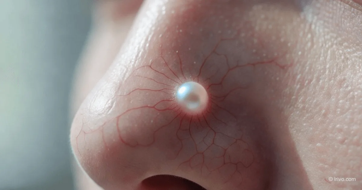
- A pearly nodule — a small, shiny, translucent or pink bump. Tiny red blood vessels (telangiectasias) are often visible through it. It looks like a small pimple that won't go away.
- A non-healing sore — a wound that bleeds, crusts over, heals, and then reappears in the same spot.
- A flat pink patch — resembles eczema or psoriasis but doesn't respond to ointments.
- A scar-like area — a whitish, waxy thickening with ill-defined borders, resembling a scar without prior injury.
BCC most commonly appears on the nose, eyelids, cheeks, forehead, and ears — areas with the most sun exposure.
2. Squamous Cell Carcinoma (SCC) — "The Scaly Aggressor"
SCC looks rougher and more "active." Distinctive features:
- A firm, scaly plaque — red or pink with a hard, rough crust.
- A wart-like growth — can grow quickly and protrude above the skin.
- A nodule with a "crater" — the center ulcerates, with raised edges.
SCC often develops from actinic keratosis — a rough, red patch that feels like sandpaper. If such a patch becomes painful, grows, or starts bleeding, the risk of progression to cancer is higher.
3. Melanoma — "The Dangerous Chameleon"
Melanoma on the face can be identified using the ABCDE rule. This is an international standard used by dermatologists worldwide:
- A (Asymmetry) — the mole is not round or oval. If you mentally divide it in half, the two halves don't match.
- B (Border) — edges are irregular, blurred, or notched, like a "moth-eaten" appearance.
- C (Color) — uneven color: a mix of brown, black, red, blue, or even white (areas of regression).
- D (Diameter) — size larger than 6 mm (about the size of a pencil eraser). However, smaller melanomas can occur on the face.
- E (Evolution) — the most important point. The mole changes: itches, bleeds, grows, or changes shape or color.
Step 2: Use the "Ugly Duckling" Method
Most people's moles and spots on the face look similar in color and shape. If you see a spot that stands out from the crowd — darker, lighter, larger, or different from your usual freckles — it's an "Ugly Duckling." Even if it doesn't fit the ABCDE rule, a doctor should see it.
Step 3: Check High-Risk Areas on the Face
Some facial areas require extra attention:
- Nose — the most common site for BCC.
- Ears and eyelids — skin here is thin, and cancer behaves more aggressively, requiring immediate treatment.
- Lip vermilion — if a scaly sore appears and doesn't heal for weeks, it could be actinic cheilitis, a precancerous condition.
- Temples and cheekbones — high-risk zones for all types of skin cancer.
Practical Tips and Important Nuances
Precancers You Should Know
Not all dangerous spots are already cancer. Some conditions are not lethal themselves but can turn into SCC or BCC.
- Actinic Keratosis (Solar Keratosis) — a red, rough, dry patch. It feels like sandpaper. Found on the face in 90% of people over 60 who have had significant sun exposure. Requires treatment (cryotherapy, laser, ointments).
- Xeroderma Pigmentosum — a rare genetic disorder where the skin cannot repair UV damage. People with this diagnosis should see a dermatologist every 3–6 months.
What to Do During a Self-Exam (per Healthline and other sources):
Perform a self-exam once a month in good lighting in front of a large mirror. Use a hand mirror to check behind the ears, under the chin, and inside the nose.
- Look for: New spots or Changes in old ones.
- Search for: Shiny nodules (BCC), Rough patches (actinic keratosis), Ulcers that don't heal for 3+ weeks.
- Assess Itching and bleeding — these are always bad signs, even if the spot is small.
Common Mistakes and How to Avoid Them
Mistake 1: "I've had this mole since childhood, so it's fine."
Even congenital nevi (moles) can transform. The rule "always been there = safe" doesn't work. What matters is the absence of change. If a mole that's "always been there" starts itching or changing color, see a doctor immediately.
Mistake 2: "I'm treating the sore with home remedies."
If a spot on your face that you think is a pimple or scratch doesn't go away in 3–4 weeks, that's not normal. Cauterizing with celandine or alcohol won't kill cancer cells; it will only make diagnosis harder by altering the tissue's appearance.
Mistake 3: "I only check for large black moles."
Melanoma isn't always black and large. There are amelanotic melanomas (pink or flesh-colored) and small melanomas (<6 mm). Relying only on "D" (Diameter) is dangerous. Focus on E (Evolution).
Summary: Key Takeaways and Next Steps
Skin cancer on the face rarely looks like a scary tumor. Usually, it's a small "deceiver": a pimple without pus, a shiny mole, or a rough patch.
What you need to do right now:
- Recall any spots you've dismissed as a "pimple" in recent months. Did it go away within 2 weeks? If not, mark that spot.
- Check your nose, forehead, and ears with a mirror and a magnifying glass.
- If you find a NEW spot (that wasn't there 6 months ago) or a CHANGE (an old spot growing/itching), schedule an appointment with a dermatologist. The doctor will perform a dermoscopy (examination with 10x magnification). It's painless and not scary.
- Don't panic. 90% of visits to a dermatologist for a "strange spot" on the face turn out to be benign. But it's better to check 10 times and hear "nothing to worry about" than to miss an early stage of cancer.
This article is based on clinical guidelines for dermato-oncology 2025–2026, data from the International Immunosuppression and Transplant Skin Cancer Collaborative, and publications from the U.S. National Library of Medicine (NIH/PubMed).
— Editorial Team